
The Baker Institute Microscopy Platform enables researchers to perform leading-edge microscopy to advance the treatment of cardiovascular disease, diabetes, and related disorders. As an open-access facility, the Baker Institute Microscopy Platform aims to advance optical imaging throughout the Baker Institute and surrounding precinct.
The Platform houses a wide range of equipment and staff expertise to enable brightfield and fluorescent imaging experiments. The Platform can assist and lead research teams across all facets of optical imaging from providing education, assisted imaging through to the development of analysis pipelines.
Platform houses state of the art equipment enable:
- live cell confocal imaging
- live intravital mouse imaging
- super-resolution imaging
- dedicated Advanced Image Analysis Workstations and software solutions.
 For more information, contact:
Adam Parslow
Microscopy Manager
T: (03) 8532 1580
E: adam.parslow@baker.edu.au
Our advanced imaging technologies
Nikon A1r Plus si NIR Modified — inverted confocal microscope
Hardware
- Observation modes: Brightfield, Fluorescence, Phase.
- Fluorescence Filter Sets for eyepiece: UV, FITC, TRITC.
- Visible Laser Lines: 405nm, 488nm, 561nm, 640nm.
- Near IR Laser Lines: 730nm, 785nm.
Research capabilities
- Integrated temperature and CO2 control for live cell imaging.
- Conventional or fast imaging (30 fps, faster with small frames).
- Optional Piezo Z-stage for rapid volume imaging.
- Simultaneous imaging with photo-activation or bleaching
- Tiling and stitching.
- 32 channel spectral detector.
- Perfect Focus System.
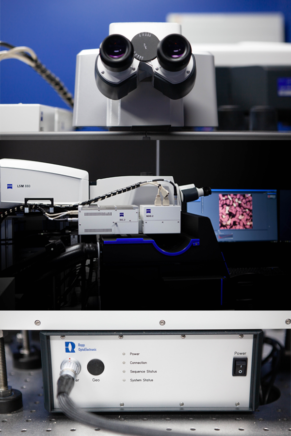
Zeiss LSM880 Airyscan — upright multiphoton microscope
Hardware
- Observation Modes: Brightfield, Fluorescence.
- Fluorescence Filter Sets for eyepiece: UV, FITC, TRITC.
- Visible Laser Lines: 405nm, 458nm, 488nm, 514nm, 561nm, 633nm.
- Infrared Laser Lines: Mai-Tai 700-1000nm & Insight X3 1050-1300nm.
- Laser Ablation System: 355nm.
- 6x external (NDD) detectors including:
- 2x external MA-PMTs and 2x GaAsP PMTs in cascade.
- Ultra-sensitive external multiphoton detection with 2x Nosepiece GaAsP detection.
Research capabilities
- Deep tissue live intravital microscopy.
- Integrated temperature incubation system.
- 32x channel spectral array for spectral linear un-mixing.
- Airyscan module for super-resolution imaging (120nm XY, 350nm Z resolution).
- Fast Airyscan imaging mode.
- Objectives for imaging thick cleared tissue.
- Tiling and stitching.
Olympus BX61 — upright motorised microscope
Hardware
- Observation Modes: Brightfield, Fluorescence, Phase, DIC.
- Fluorescence Filter Sets: DAPI; FITC; TRITC; Cy5; Cy5.5.
- Camera: Olympus DP80. Dual CCD Monochrome and Colour Camera.
Research capabilities
- Fully motorised microscope for multi-channel acquisition.
- Polarised light microscopy.
Image analysis workstation
Hardware
- HP Z6 Workstation.
- Processor: Intel Xeon Gold 6134.
- Memory: 192 GB (6x32GB) DDR4-2666 MHz ECC registered RAM.
- Graphics card: NVIDIA Quadro P4000 8GB DP.
- Hard drive: 8TB RAID10.
Software
- Imaris 9.9.
- HALO and HALO AI.
- Arvis 3.2.
- FIJI.
- QuPath.
- Rstudio.
- Zen Blue 2.3.
- Zen Black 2.3
Location
Baker Heart and Diabetes Institute
75 Commercial Road, Melbourne
Victoria 3004, Australia