
Cardiac Cellular Systems





About the Cardiac Cellular Systems laboratory
Heart failure is a leading cause of death worldwide. It arises from many different insults — heart attack, high blood pressure, diabetes and obesity among them — and is characterised by harmful cardiac remodelling and declining heart function, ultimately leading to organ failure and death. There is still no cure for heart failure, and fundamental questions remain about how cardiac remodelling occurs and whether it can be reversed.
Our laboratory works at the level of the heart's cell network. We map the cells of the heart, define the genetic programs that switch them into disease-driving states, and are now developing new medicines designed to rewire those cells to repair the heart and prevent heart failure.
Discovering the heart's cellular ecosystem
Until recently, the cellular composition of the heart was poorly understood. Using advanced genetic, flow cytometric and single-cell transcriptomic approaches, our laboratory has shed new light on what the heart is actually made of. We've demonstrated that the heart comprises a complex and diverse ecosystem of non-myocytes — the cells that support and surround heart muscle cells (Figure 1: Skelly et al. 2018, Pinto et al. 2016, Pinto et al. 2012).
We have since shown that this ecosystem is not fixed. It is remodelled by disease, with specific scar-forming fibroblast populations emerging during cardiac stress that are also found in human heart disease (McLellan et al. 2020), and it is shaped by biological sex and gonadal hormones (Squiers et al. 2021). Most recently, we have moved from describing these cell states to defining the transcription factor circuits that create them, mapping the gene-regulatory programs that establish and maintain a scar-forming identity across both mouse and human hearts (Krstevski et al. 2026).
Knowing which cells drive disease, and which molecular switches control them, makes it possible to target and manipulate specific cell types deliberately — and that's where our research now comes in.
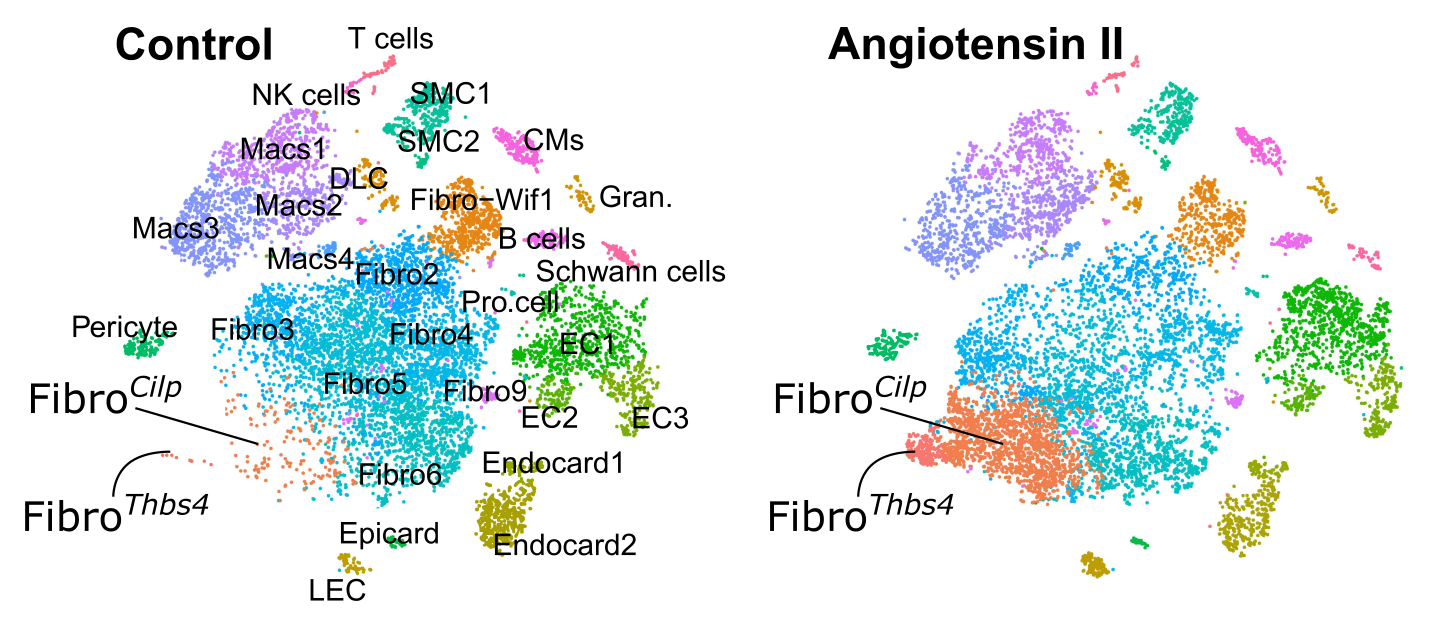
Figure 1: Understanding cardiac cellular diversity and phenotypes in tissue homeostasis and stress. The figure shows tSNE plots with dots representing individual cells. Fibro-Cilp and Fibro-Thbs4 that emerge after angiotensin II-induced fibrosis are indicated from McLellan et al. 2020.
Our research aims
We have two overarching goals:
- Understand how cardiac cell networks contribute to heart failure
We're determining how pathological remodelling of the cardiac cell network drives heart failure — after ischaemic injury, in diabetes and hypertension, and in response to emerging environmental stressors such as micro- and nanoplastics. - Rewire cardiac cell networks to prevent and treat heart disease
We're developing ways to intervene in that network — including programmable RNA medicines, small molecules and endocrine factors — to halt harmful remodelling and convert scar formation into genuine heart repair (Figure 2).
Using single-cell and multiomic technologies, data science, micro-anatomy, gene editing and traditional mouse genetics, our research takes an integrative systems biology approach to uncovering the fundamental processes that govern cell networks in the heart, and to turning that understanding into therapies.
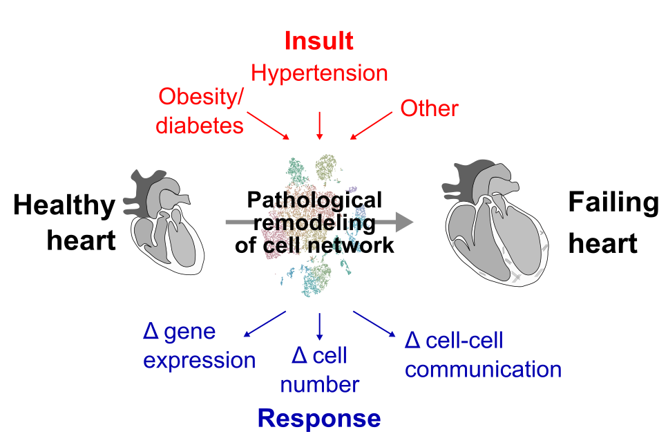
Figure 2: The cardiac cell network as a therapeutic target. Cardiac insults — ischaemic injury, diabetes and hypertension, and novel environmental stressors — drive pathological remodelling of the cardiac cell network (red), converting a healthy heart into a failing heart. Our work aims to interrupt and reverse this process using targeted interventions (blue), including small molecules, mRNA therapeutics delivered by lipid nanoparticles, and endocrine factors.
Key areas of ongoing research
- The regulatory logic of scar-forming cells
Defining the transcription factor circuits that give scar-forming fibroblasts their identity after a heart attack, using paired single-cell transcriptomic and epigenomic profiling of mouse and human heart tissue, to identify regulators that can be targeted therapeutically. - Turning scar into muscle
Discovering and optimising the factors that can reprogram scar-forming fibroblasts into cardiomyocyte-like cells, and developing gene-activation strategies that make this switch durable. - Targeted RNA medicines for the heart
Engineering lipid nanoparticles and 'smart' mRNA designs that deliver therapeutic instructions selectively to scar-forming cells in the injured heart, while limiting activity in off-target tissues — a scalable, non-viral alternative to viral gene therapy. - Cardiac cell network plasticity and resilience
Determining how cardiac cell networks respond and adapt to physiological and environmental stressors, including obesity, diabetes, hypertension, and exposure to micro- and nanoplastics. - Novel research tools and open data
Developing genetic, computational and imaging approaches to study diverse cell populations precisely, and sharing them with the field — including CLARA, our Cardiovascular Atlas (clara.baker.edu.au), our open-access portal for exploring the cardiovascular cellular landscape, now used by researchers in more than 25 countries.
Key areas of technical expertise
- Single-cell and multiomic biology
Including single-cell RNA sequencing, paired single-cell transcriptomic and epigenomic (multiomic) profiling, high-dimensional flow cytometry and image cytometry. - Computational biology and data science
Including gene-regulatory network inference, cross-species comparative analysis and interactive data sharing through CLARA. - RNA therapeutics and nanoparticle delivery
Including mRNA design, cell-selective 'smart' mRNA, and high-throughput screening of lipid nanoparticle formulations for cardiac cell targeting. - Gene editing
Including CRISPR activation and prime editing approaches for durable control of gene expression. - Multidimensional imaging
Including 3D imaging and spatial mapping of cellular interactions. - Mouse genetics
Including development of novel cell- and organ-specific genetic tools and intersectional lineage-tracing systems for tracking cell fate after injury.
We work closely with colleagues across the Baker Institute — including the Single-Cell Omics Platform, the Centre for Cardiometabolic mRNA Therapy, the CRISPR Validation Platform and the Translational Cardiology Centre — and with national and international collaborators in nanoparticle chemistry, gene editing and human cardiac tissue research.
Community engagement
We collaborate with the Baker Institute's Community Engagement Group to embed meaningful consumer and community voices into our research. This engagement has helped shape our study direction, refine recruitment and messaging, and support more relevant, accessible and impactful outcomes for people affected by heart failure and cardiac disease. We also work with heart attack survivors and community members to keep our therapeutic goals aligned with what matters most to patients, and our team contributes regularly to public discussion of heart health and emerging cardiovascular risks.
Key publications
- Conserved transcriptional circuits regulate cardiac fibroblast-mediated fibrosis
Circulation Research 2026 - Spatio-temporal targeting of cardiac cells with lipid nanoparticles after myocardial infarction
Journal of Molecular and Cellular Cardiology Plus 2026 - Cardiac cellularity is dependent upon biological sex and is regulated by gonadal hormones
Circulation Research 2021 - High-resolution transcriptomic profiling of the heart during chronic stress reveals cellular drivers of cardiac fibrosis and hypertrophy
Circulation 2020 - Single-cell transcriptional profiling reveals cellular diversity and intercommunication in the mouse heart
Cell Reports 2018

There's a way to give that's right for you
Explore ways to give
Whether you make a one-off gift, give regularly or leave a lasting legacy, your support helps us keep doing research that saves lives.