27 May 2025
Media release

The Baker Heart and Diabetes Institute is establishing Australia’s first imaging facility to examine energy levels and fat accumulation in the heart muscle. This will identify the need for personalised treatments to reverse and prevent heart disease associated with obesity, diabetes and related diseases.
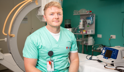
Our new cardiac energetics imaging facility is led by Professor Eylem Levelt, a researcher and cardiologist from the UK who joined the Institute earlier this year. Her expertise, embracing the latest in heart imaging techniques, will form part of our Centre of Cardiometabolic Imaging and Obesity.
Professor Levelt is using MR Spectroscopy — molecular level imaging — to monitor energetics and fat accumulation in the heart. The scans for energy assessment of the heart take just 12 minutes and similar scans for measuring fat accumulation in the heart take 5 minutes. Both techniques are non-invasive and require no radiation or injections.
She is also utilising the latest developments in MRI techniques to measure blood supply and oxygenation of the heart muscle. In combination, these sensitive techniques allow us to comprehensively evaluate the earliest subtle abnormalities in the heart which contribute to development of heart disease over time.
People who are overweight or living with obesity are at high risk of developing heart failure and at risk of premature death related to heart failure. By examining the energy levels and accumulation of fat within the heart, we hope to identify those on the obesity trajectory before conditions like heart failure set in.
Professor Levelt says assessing the energetic state of the heart is one of the most sensitive monitoring tools. A decrease in energetics signals that something is not right and can be used as a diagnostic tool to identify people who are at risk of developing heart failure complications from diabetes and obesity.
She says compromised energy production in the mitochondria is a key contributor to heart disease in type 2 diabetes and obesity. The molecule carrying energy within the cells is known as ATP, or adenosine triphosphate, which is an energy currency. Mitochondria provide the bulk of the ATP needed for heart muscle contraction. The heart uses about six kilograms of ATP a day, while it contains very little ATP storage, so the mitochondria have to work continuously to produce it. Our technique is indirectly informing us about energy production in the heart.
Professor Levelt says: “The heart is an energy-expensive organ, using more energy than its total weight every day, and that’s why energy metabolism is critical for maintaining normal function of the heart. Using advanced imaging techniques allows a view of the chemistry of the heart muscle, revealing its energy management. If we can detect inefficient energy management with our sensitive tools, we can take the necessary precautions to improve myocardial energy production and reduce the risk of heart failure development.”
For further information or to organise interviews please contact:
Loretta Walshe
T: 03 8532 1240
M: 0466 941 942
E: loretta.walshe@baker.edu.au